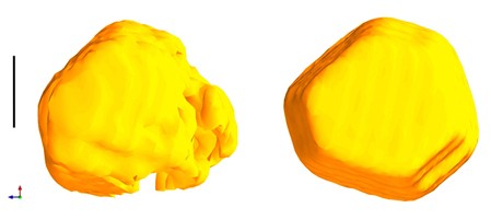
Shown on the left is the three dimensional image
of a gold nanocrystal obtained previously while on the right is the image using
the newly developed method. The features
of the nanocrystal are vastly improved in the image on the right. The black scale bar is 100 nanometres (1
nanometre = 1 billionth of a meter).
A new advance in X-ray imaging has revealed the dramatic three-dimensional shape of gold nanocrystals, and is likely to shine a light on the structure of other nano-scale materials. Described today in Nature , the new technique improves the quality of nanomaterial images, made using X-ray diffraction, by accurately correcting distortions in the X-ray light. Jesse Clark, lead author of the study from the London Centre for Nanotechnology said: "With nanomaterials playing an increasingly important role in many applications, there is a real need to be able to obtain very high quality three dimensional images of these samples. "Up until now we have been limited by the quality of our X-rays. Here we have demonstrated that with imperfect X-ray sources we can still obtain very high quality images of nanomaterials." Up until now, most nanomaterial imaging has been done using electron microscopy. X-ray imaging is an attractive alternative as X-rays penetrate further into the material than electrons and can be used in ambient or controlled environments. However, making lenses that focus X-rays is very difficult.
TO READ THIS ARTICLE, CREATE YOUR ACCOUNT
And extend your reading, free of charge and with no commitment.