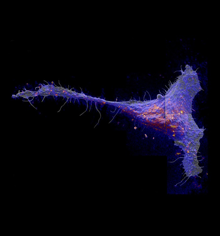
Researchers found that a class of molecules called sphingolipds congregate in large patches in the cell membrane. Red and yellow colors indicate local elevations in the sphingolipid abundance.
CHAMPAIGN, Ill. Sight would dramatically alter a blind man's understanding of an elephant, according to the old story. Now, a look directly at a cell surface is changing our understanding of cell membrane organization. Using a completely new approach to imaging cell membranes, a study by researchers from the University of Illinois, Lawrence Livermore National Laboratory and the National Institutes of Health revealed some surprising relationships among molecules within cell membranes. Led by Mary Kraft , a U. of I. professor of chemical and biomolecular engineering , the team published its findings in the Proceedings of the National Academy of Sciences. Cells are enveloped in semi-permeable membranes that act as a barrier between the inside and outside of the cell. The membrane is mainly composed of a class of molecules called lipids, studded with proteins that help regulate how the cell responds to its environment.
TO READ THIS ARTICLE, CREATE YOUR ACCOUNT
And extend your reading, free of charge and with no commitment.