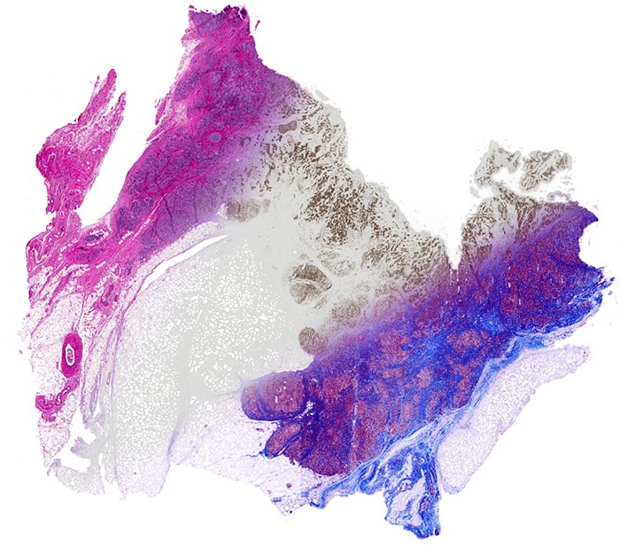
Breast tissue is computationally stained using data from infrared imaging without actually staining the tissue, enabling multiple stains on the same sample. From left, the image shows a Hematoxylin and Eosin stain (pink-blue), molecular staining for epithelial cells (brown color) and Masson’s trichrome (blue, red at right).
CHAMPAIGN, Ill. One infrared scan can give pathologists a window into the structures and molecules inside tissues and cells, enabling fast and broad diagnostic assessments, thanks to an imaging technique developed by University of Illinois researchers and clinical partners. Using a combination of advanced microscope imaging and computer analysis, the new technique can give pathologists and researchers precise information without using chemical stains or dyes. Led by Rohit Bhargava , U. of I. professor of bioengineering and member of the Beckman Institute for Advanced Science and Technology , the researchers published their findings in the journal Technology. "Any sample can be analyzed for desired stains without material cost, time or effort, while leaving precious tissue pristine for downstream analyses," Bhargava said. To study tissue samples, doctors and researchers use stains or dyes that stick to the particular structure or molecule they are looking for. Staining can be a long and exacting process, and the added chemicals can damage cells.
TO READ THIS ARTICLE, CREATE YOUR ACCOUNT
And extend your reading, free of charge and with no commitment.