University of Illinois engineers developed a method to computationally correct aberrations in three-dimensional tissue microscopy.
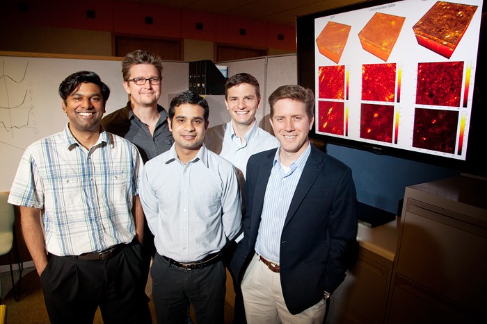
From left, postdoctoral researcher Steven Adie, P. Scott Carney, graduate students Adeel Ahmad and Benedikt Graf, and Stephen Boppart.
University of Illinois engineers developed a method to computationally correct aberrations in three-dimensional tissue microscopy. From left, postdoctoral researcher Steven Adie, professor P. Scott Carney, graduate students Adeel Ahmad and Benedikt Graf, and professor Stephen Boppart. Photo by L. Brian Stauffer CHAMPAIGN, Ill. Real-time, 3-D microscopic tissue imaging could be a revolution for medical fields such as cancer diagnosis, minimally invasive surgery and ophthalmology. University of Illinois researchers have developed a technique to computationally correct for aberrations in optical tomography, bringing the future of medical imaging into focus. Aberrations in imaging can make points appear as slashes or blurs. Click on image to see image corrected by computational adaptive optics developed by U. of I. researchers.
TO READ THIS ARTICLE, CREATE YOUR ACCOUNT
And extend your reading, free of charge and with no commitment.