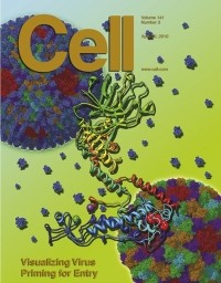
Artistic representation of a Cryo-EM 3-D reconstruction of aquareovirus
UCLA researchers report in the April 30 edition of the journal Cell that they have imaged a virus structure at a resolution high enough to effectively "see" atoms, the first published instance of imaging biological complexes at such a resolution. The research team, led by Hong Zhou, UCLA professor of microbiology, immunology and molecular genetics, used cryo-electron microscopy to image the structure at 3.3 angstroms. An angstrom is the smallest recognized division of a chemical element and is about the distance between the two hydrogen atoms in a water molecule. The study, the researchers say, demonstrates the great potential of cryo-electron microscopy, or Cryo-EM, for producing extremely high-resolution images of biological samples in their native environment. "This is the first study to determine an atomic resolution structure through Cryo-EM alone," said Xing Zhang, a postdoctoral candidate in Zhou's group and lead author of the Cell paper. "By proving the effectiveness of this microscopy technique, we have opened the door to a wide variety of biological studies." With traditional light microscopy, a magnified image of a sample is viewed through a lens. Some samples, however, are too small to diffract visible light (in the 500 to 800 nm range, or 5,000 to 8,000 angstroms) and therefore cannot be seen.
TO READ THIS ARTICLE, CREATE YOUR ACCOUNT
And extend your reading, free of charge and with no commitment.