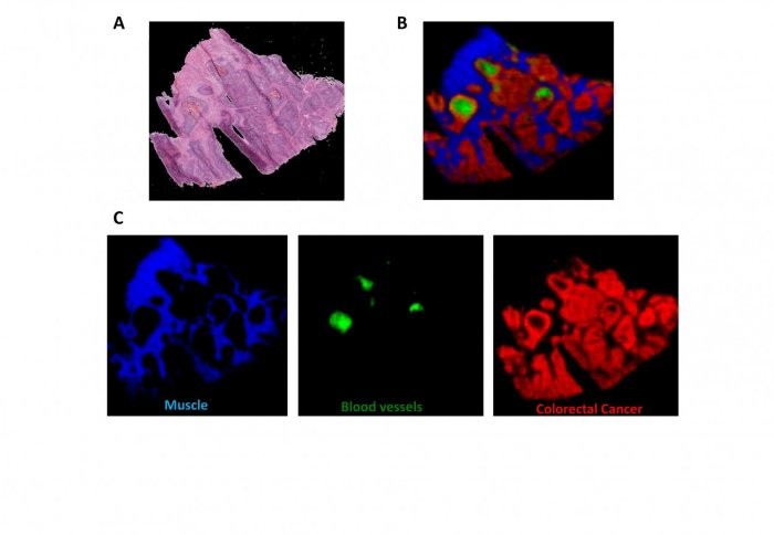
A section of bowel tissue as an optical image (A) and using mass spectrometry imaging to identify tissue types (B and C).
A new method for analysing biological samples based on their chemical makeup is set to transform the way medical scientists examine diseased tissue. When tests are carried out on a patient's tissue today, such as to look for cancer, the test has to be interpreted by a histology specialist, and can take weeks to obtain a full result. Mass spectrometry imaging (MSI) uses technologies that reveal how hundreds or thousands of chemical components are distributed in a tissue sample. Scientists have proposed using MSI to identify tissue types for many years, but until now, no method has been devised to apply such technology to any type of tissue. In this week's Proceedings of the National Academy of Sciences , researchers at Imperial College London have outlined a recipe for processing MSI data and building a database of tissue types. In MSI, a beam moves across the surface of a sample, producing a pixelated image. Each pixel contains data on thousands of chemicals present in that part of the sample.
TO READ THIS ARTICLE, CREATE YOUR ACCOUNT
And extend your reading, free of charge and with no commitment.