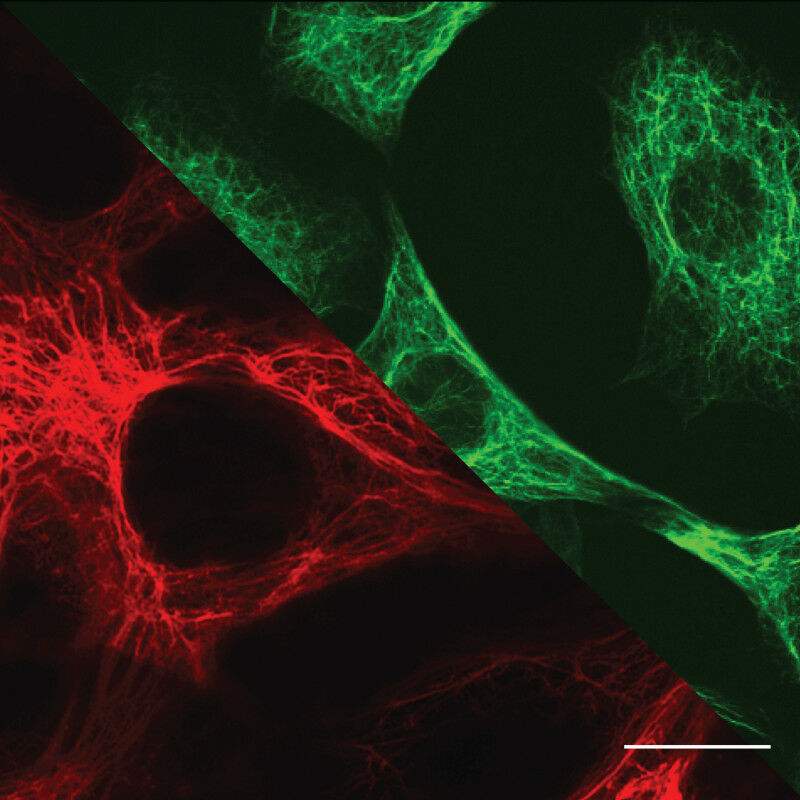
Microscopy images of biological cells: top right (green) - Vimentin intermediate filaments in fibroblasts; bottom left (red) - Keratin intermediate filaments in epithelial cells. Scale: 10 µm. Photo: top right (green): Ulrike Rölleke. bottom left (red): Ruth Meyer
Microscopy images of biological cells: top right ( green ) - Vimentin intermediate filaments in fibroblasts; bottom left ( red ) - Keratin intermediate filaments in epithelial cells. Scale: 10 µm. Photo: top right ( green ): Ulrike Rölleke. bottom left ( red ): Ruth Meyer Research team at Göttingen University discovers surprising properties of the cytoskeleton Most biological cells have a fixed place in an organism. However, cells can become mobile and move through the body. This happens, for example, during wound healing or when tumour cells divide uncontrollably and migrate through the body. Mobile and stationary cells differ in various ways, including their cytoskeleton.
TO READ THIS ARTICLE, CREATE YOUR ACCOUNT
And extend your reading, free of charge and with no commitment.