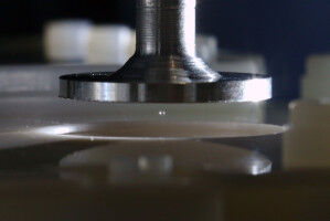
EPFL scientists have developed a scanning transmission electron microscopy method that can quickly and efficiently generate 3D representations of curvilinear nanostructures. Image caption: Superposed, tilt-less electron microscopy stereo image (color-filtered) of carbon nanospheres decorated with nanoparticles. The same structures appear in red and blue and the nanoparticles are slightly shifted according to their 3D distribution in the carbon sphere. This image shows the applicability of the new tilt-less 3D imaging techniques to other structures. Physical and biological sciences increasingly require the ability to observe nano-sized objects. This can be accomplished with transmission electron microscopy (TEM), which is generally limited to 2D images. Using TEM to reconstruct 3D images instead usually requires tilting the sample through an arc to image hundreds of views of it and needs sophisticated image processing to reconstruct their 3D shape, creating a number of problems.
TO READ THIS ARTICLE, CREATE YOUR ACCOUNT
And extend your reading, free of charge and with no commitment.
Your Benefits
- Access to all content
- Receive newsmails for news and jobs
- Post ads