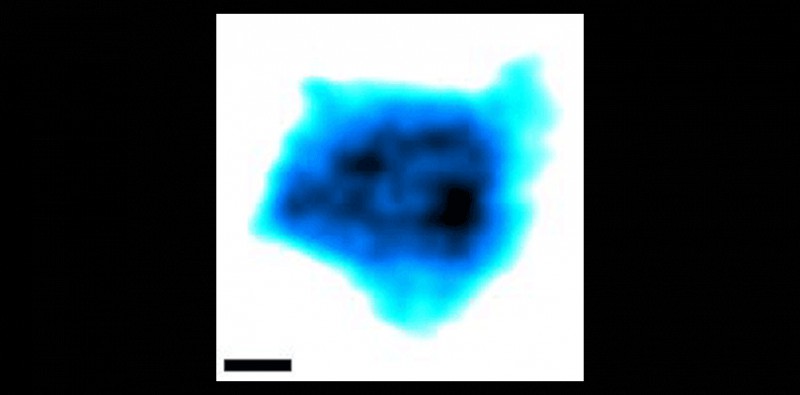
Video imaging by high-speed AFM captures native nuclear pore complexes at work; the inset scale bar is 10 nanometers. (Image: University of Basel)
Using an ultra fast-scanning atomic force microscope, a team of researchers from the University of Basel has filmed "living" nuclear pore complexes at work for the first time. Nuclear pores are molecular machines that control the traffic entering or exiting the cell nucleus. In their article published , the researchers explain how the passage of unwanted molecules is prevented by rapidly moving molecular "tentacles" inside the pore. The atomic force microscope (AFM) is not a microscope to look through. Like a blind man uses his fingers, it "feels" a surface with an extremely fine tip to resolve tiny cellular structures of only millionths of a millimeter in size, such as the pores in the nuclear envelope. However, this process is normally slow and can take up to one minute to capture an image. In comparison, modern high-speed AFMs are able to record movies of molecules in action by capturing several hundred images per minute.
TO READ THIS ARTICLE, CREATE YOUR ACCOUNT
And extend your reading, free of charge and with no commitment.