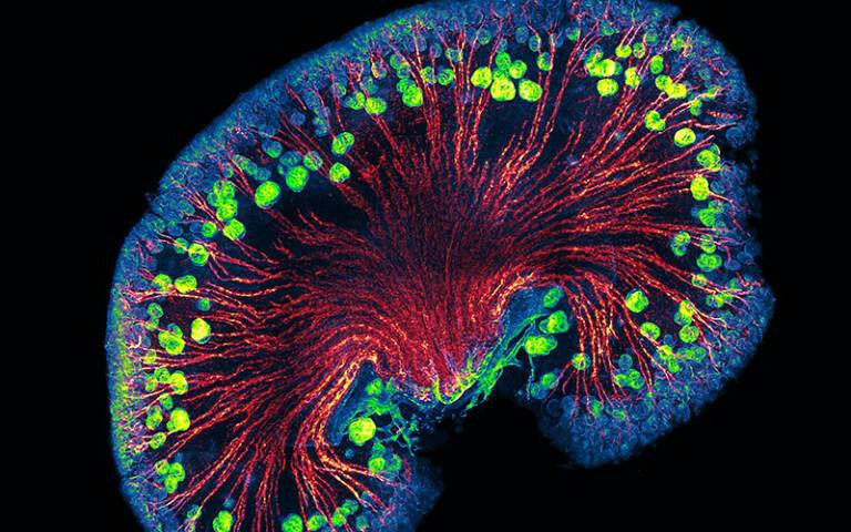
A confocal microscopy image of the urinary filtration apparatus of the developing kidney. A confocal microscopy image of the urinary filtration apparatus of the developing kidney. Credit: David Long, Claire Walsh and Danyal Jafree.
A confocal microscopy image of the urinary filtration apparatus of the developing kidney. A confocal microscopy image of the urinary filtration apparatus of the developing kidney. Credit: David Long, Claire Walsh and Danyal Jafree. Interdisciplinary team from UCL recognised with prestigious photography prize for their work to develop new techniques to view the internal workings of human organs. Professor David Long, Dr Daniyal Jafree (UCL Great Ormond Street Institute of Child Health) and Dr Claire Walsh (UCL Mechanical Engineering) collected the Combined Royal Colleges Medal for their advances in imaging to better understand kidney disease. Using a revolutionary technology called Hierarchical Phase-Contrast Tomography (HiP-CT) and through the Human Organ Atlas project , the interdisciplinary team of engineers, biologists and clinicians made it possible to view the entire human kidney in three dimensions. This allowed them to zoom down to the level of individual kidney filters and vessels.
TO READ THIS ARTICLE, CREATE YOUR ACCOUNT
And extend your reading, free of charge and with no commitment.