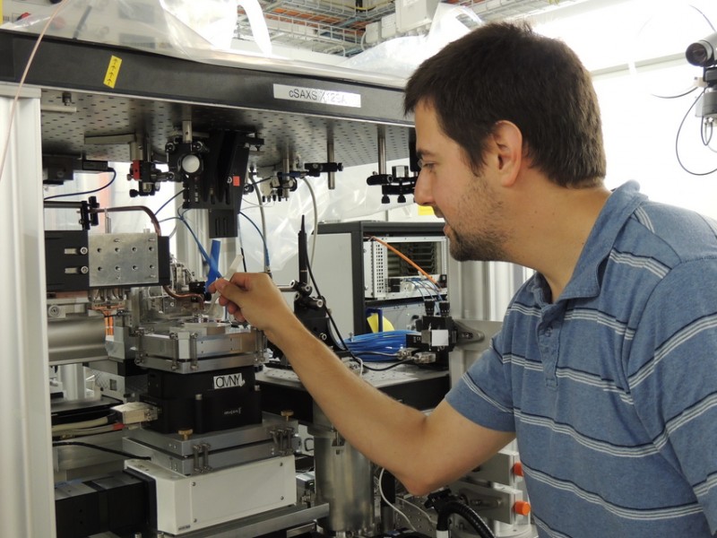
Mirko Holler fixing a sample to the measuring unit for ptychographic tomography at the SLS.
Media Releases Research Using Synchrotron Light World-record resolution in tomography with hard X-rays Tomography enables the interior of a vast range of objects to be depicted in 3D - from cellular structures to technical appliances. Researchers from the Paul Scherrer Institut (PSI) have now devised a method that opens up new scales of tomographic imaging and will thus make the detailed study of representative volumes of biological tissue and materials science specimens possible in future. Until now, the relevant details on a scale of a few nanometres were only visible with methods that required very thin samples. With the aid of a special prototype set-up at the PSI's Swiss Light Source (SLS) the researchers have now achieved a 3D resolution of sixteen nanometres on a nanoporous glass test sample, a feat that is unmatched for X-ray tomography. The measurement is non-destructive, so it allows to study small details in the context of their surroundings or to analyse larger sample volumes in such a way that the information obtained is influenced less by locally induced variances. The resolution of 16 nm was achieved on a prototype of the OMNY instrument, which is still under construction. The final version will enable the researchers to cool down the sample during the experiment to prevent X-ray induced sample damage.
TO READ THIS ARTICLE, CREATE YOUR ACCOUNT
And extend your reading, free of charge and with no commitment.