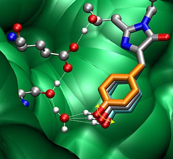
After GFP’s chromophore absorbs a blue photon, its excited phenoxyl-ring wags rapidly back and forth, settling into a position that allows a positively charged hydrogen atom – a bare proton – to hop along the dotted lines, leading to bright green fluorescence. The red balls are oxygen atoms, the small silver balls are hydrogen atoms (protons), the large silver balls are carbon atoms, and the blue balls are nitrogen. The green background is the barrel-like structure of GFP, which encloses the central chromophore.
BERKELEY — University of California, Berkeley, chemists have discovered the secret to the success of a jellyfish protein whose green glow has made it the darling of biologists and the subject of the 2008 Nobel Prize in Physiology or Medicine. After GFP's chromophore absorbs a blue photon, its excited phenoxyl-ring wags rapidly back and forth, settling into a position that allows a positively charged hydrogen atom - a bare proton - to hop along the dotted lines, leading to bright green fluorescence. The red balls are oxygen atoms, the small silver balls are hydrogen atoms (protons), the large silver balls are carbon atoms, and the blue balls are nitrogen. The green background is the barrel-like structure of GFP, which encloses the central chromophore. (Renee Frontiera & Chong Fang/UC Berkeley) GFP has replaced many dyes in biological studies because it is non-toxic and, when attached to a gene and inserted into an organism, serves as a bright, glowing confirmation that the gene has hit its target. Obtained originally from a bioluminescent Pacific Ocean jellyfish, the protein has been mutated and engineered to absorb and emit various colors. The UC Berkeley chemists used extremely short laser pulses 20 millionths of a nanosecond, or 20 femtoseconds to take snapshots of GFP to determine the structural changes it undergoes when it fluoresces.
TO READ THIS ARTICLE, CREATE YOUR ACCOUNT
And extend your reading, free of charge and with no commitment.