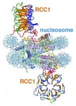
This image illustrates the RCC1 chromatin protein interacting with the nucleosome. A high-resolution file of this image and a video animation are online at
University Park, Pa. — In a landmark study to be published , scientists have been able to create the first picture of genetic processes that happen inside every cell of our bodies. Using a 3-D visualization method called X-ray crystallography, Song Tan , associate professor of biochemistry and molecular biology at Penn State, has built the first-ever image of a protein interacting with the nucleosome - DNA packed tightly into space-saving bundles organized around a protein core. The research is expected to aid future investigations into diseases such as cancer. As the genetic blueprint of life, DNA must be deciphered or "read," even when densely packed into nucleosomes. The nucleosome is therefore a key target of genetic processes in a cell and a focus of scientific investigations into how normal and diseased cells work. Previous studies at Penn State and other research institutions led to the discovery of chromatin enzymes - proteins that act to turn specific genes on or off by binding to the nucleosome.
TO READ THIS ARTICLE, CREATE YOUR ACCOUNT
And extend your reading, free of charge and with no commitment.