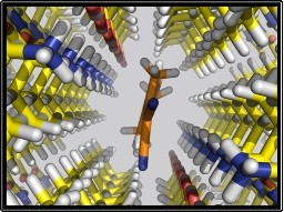
Top view: Imaging agent’s chemical composition
In the Alzheimer's brain, hard plaques accumulate between the nerve cells, while twisted fibers grow inside the nerve cells. The plaques arise from protein fragments called beta amlyoid, and the fibers form from a protein called tau. Doctors rely on brain scans to detect amyloid and tau and provide early intervention and treatments that potentially slow or reverse disease progression. But how exactly the imaging agents work that scan the Alzheimer's brain has not been known. Now, new UCLA study reveals the physical mechanisms that allow these chemical agents to bind to and detect amyloid beta plaques and tau tangles in the brain. Kendall Houk, the Saul Winstein Professor of Organic Chemistry in UCLA's College of Letters and Sciences, and Jorge Barrio, distinguished professor of molecular and medical pharmacology at the David Geffen School of Medicine at UCLA, are available for s.
TO READ THIS ARTICLE, CREATE YOUR ACCOUNT
And extend your reading, free of charge and with no commitment.