Long considered unrealistic, electron microscopy-based connectomics has established itself as a revolution in neuroscience. In 2025, the journal Nature Methods named it Method of the Year, recognizing a technology capable of reconstructing neural circuits in three dimensions with nanometric resolution. At the Friedrich Miescher Institute for Biomedical Research (FMI) in Basel, Rainer Friedrich’s laboratory and specialists such as Alexandra Graff Meyer have played a pioneering role in the development of this approach, which links the brain’s architecture to its functioning.
Thanks to these advances, scientists are closer than ever to understanding the mechanisms by which neural networks produce perception, memory and intelligence.
An idea once thought unfeasible
In the early 2000s, few neuroscientists would have bet that electron microscopy could one day map entire neural circuits in three dimensions. The technique was slow, complex and provided only very thin slices of brain tissue, with no overall view of the connections underlying thought and memory.
Twenty years on, the situation has changed radically. Today, connectomics using electron microscopy enables complete neural networks to be reconstructed with unrivalled precision, transforming the way researchers study the brain.
Linking brain structure to function
Among the researchers who have contributed to this transformation is Rainer Friedrich, one of the pioneers of the field. At FMI, his laboratory helped transform connectomics from a speculative idea into a powerful experimental approach, capable of establishing a direct link between neural wiring and brain function.
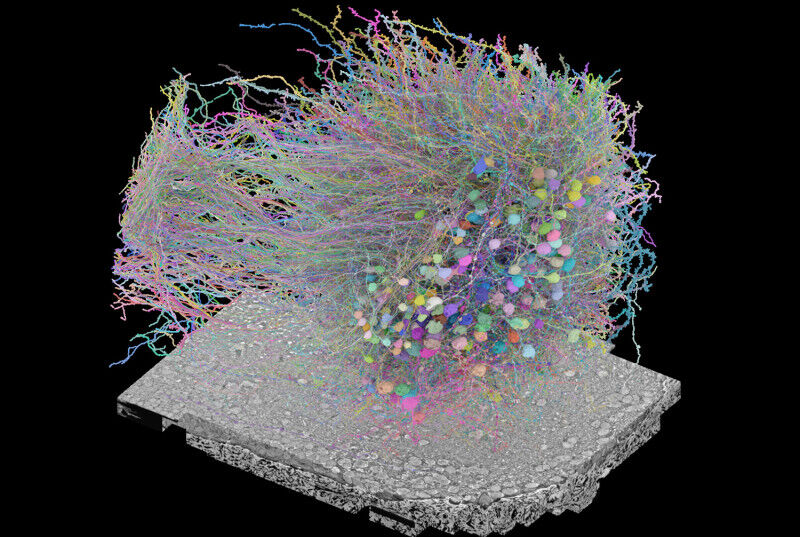
The method involves reconstructing neural circuits at nanometer scale using three-dimensional electron microscopy, making it possible to track each neuron individually and map all the synapses that link them.
"It’s in the connections that function lies," explains Friedrich. For decades, neuroscientists have tried to understand how the brain works without being able to observe its complete wiring, as if we were trying to dismantle a machine without seeing its internal circuits. Now, connectomics enables us to directly confront theoretical models with biological reality.
A pioneering path
Friedrich’s interest in connectomics dates back to his early days at the Max Planck Institute for Medical Research in Heidelberg, where he worked alongside physicist Winfried Denk, one of the inventors of modern serial electron microscopy techniques.
"We tested these new methods together to answer neuroscience questions, often late at night", Friedrich recalls. "At the time, it was not at all obvious that the technique could be used on a large scale. "
Later, at the IMF, he decided to invest fully in this approach. Thanks to access to specialized equipment and close collaboration with the electron microscopy center shared with Novartis, the method is rapidly evolving. The FMI’s environment, combining neurophysiology, computational neuroscience and advanced microscopy, is conducive to these advances.
Understanding how neurons produce intelligence
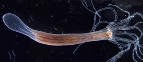
Friedrich’s team studies how neural networks give rise to memory, perception and intelligent behavior. In particular, the researchers are working on zebrafish, focusing on the sense of smell and how the brain constructs internal representations of the environment.
In a landmark study, the team showed how a network of over a thousand neurons in the olfactory bulb - the first odor processing station - transforms very similar sensory signals into distinct patterns of activity. This mechanism enables the animal to learn and distinguish between almost identical odors.
"This type of calculation is fundamental to many learning processes," explains Friedrich. "But it was impossible to understand it without knowing precisely the connections between neurons. "
Thanks to connectomics, the researchers were able to reconstruct all the connections in the olfactory bulb of a larval zebrafish and show how the organization of the network explains the observed functioning.
A technology based on teamwork
Progress in connectomics relies on the coordinated work of numerous specialists. At the FMI, neuroscientists work closely with experts responsible for preparing samples, carrying out imaging and developing the necessary IT tools.
Technical specialist Alexandra Graff Meyer prepares samples, participates in imaging experiments and develops protocols for reliable mapping of neural circuits.
In connectomics, brain tissue is imaged layer by layer at very high resolution, then thousands of images are assembled to reconstruct a three-dimensional map of connections. But the work begins long before microscopy.
In Friedrich’s laboratory, experiments begin by studying the behavior of zebrafish larvae and calcium imaging, which records the activity of thousands of neurons live. These data are used to identify the circuits to be analyzed in detail.
25-nanometer slices
Preparing samples for electron microscopy can take up to a week. The tissue is fixed to preserve its structure, stained to create contrast, then embedded in a resin to obtain a block strong enough to be cut into ultra-thin slices.
using a microtome fitted with a diamond knife, sections just 25 nanometers thick are cut out, some four thousand times thinner than a human hair.
"It’s like cutting carpaccio," explains Graff Meyer. "If a single slice is lost, months of work can be compromised, as the continuity of neuronal connections is broken. "
Millions of images to reconstruct the brain
To automate this process, the researchers developed SBEMimage, an open source software package that automatically acquires serial electron microscopy images. Acquisitions can take several days, or even weeks.
But imaging is only the first step. Millions of images must then be aligned, reconstructed and analyzed using computer tools capable of transforming the raw data into navigable three-dimensional maps.
At the FMI, microscopy specialist Tomas Gancarcik oversees the imaging and initial data processing stages, ensuring the quality necessary for reliable reconstruction.
The final reconstruction of neural circuits is carried out by the neuroscientists themselves. "Advances come from bringing together very different skills around a single project," Friedrich emphasizes.
Towards a deeper understanding of the brain
For researchers, connectomics opens up a new era. By combining meticulous preparation, cutting-edge imaging and computer analysis, it is finally possible to observe how the organization of the brain gives rise to mental functions.
"When preparation and imaging are successful, we can begin to understand how the brain really works," concludes Alexandra Graff Meyer.