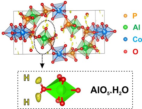
 © P. Boullay CRISMAT (CNRS/Ensicaen/Unicaen). Representation of the structure of a cobalt aluminophosphate, superimposed onto a map showing maxima (in yellow) associated with the hydrogen positions, after analysis of the electron diffraction data.
Diffraction-based analytical methods are widely used in laboratories, but they struggle to study samples that are smaller than a micrometer in size. Researchers from the Laboratoire de cristallographie et sciences des matériaux (CNRS/Ensicaen/Unicaen), the Laboratoire catalyse et spectrochimie (CNRS/Ensicaen/Unicaen)
1, and the Academy of Sciences of the Czech Republic have nevertheless been successful in using electron diffraction to reveal the structure of nanocrystals
2. Their method is so sensitive that it has even located the position of hydrogen atoms for the first time, which is crucial in accessing the morphology of the molecules or the size of cavities in porous materials. Diffraction of X-rays or neutrons by crystals is a method of choice for obtaining the atomic structure of crystalline solids essential for understanding the properties of materials, reactional mechanisms or biomolecules like proteins or DNA. However, this technique requires crystals of the order of a micrometer, in the case of X-rays, and of a millimeter, in the case of neutrons. Electron diffraction enables the study of nanosized samples, thanks to the strong interaction with the material of these charged particles. The downside is that multiple diffractions occur and reduce the quality of the results obtained.
TO READ THIS ARTICLE, CREATE YOUR ACCOUNT
And extend your reading, free of charge and with no commitment.
Your Benefits
- Access to all content
- Receive newsmails for news and jobs
- Post ads