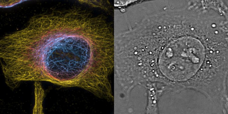
 A cell visualized with the PRISM method
In a breakthrough for biological imaging, EPFL scientists have developed the first microscope platform that can perform super-resolution spatial and temporal imaging, capturing unprecedented views inside living cells. The landmark paper is published. Super-resolution microscopy is a technique that can "see" beyond the diffraction of light, providing unprecedented views of cells and their interior structures and organelles. The technique has garnered increasing interest recently, especially since its developers won the Nobel Prize in Chemistry in 2014. But super-resolution microscopy comes with a big limitation: it only offers spatial resolution. That might suffice for static samples, like solid materials or fixed cells, but when it comes to biology, things become more complicated. Living cells are highly dynamic and depend on a complex set of biological processes that occur across subsecond timescales, constantly changing.
TO READ THIS ARTICLE, CREATE YOUR ACCOUNT
And extend your reading, free of charge and with no commitment.
Your Benefits
- Access to all content
- Receive newsmails for news and jobs
- Post ads