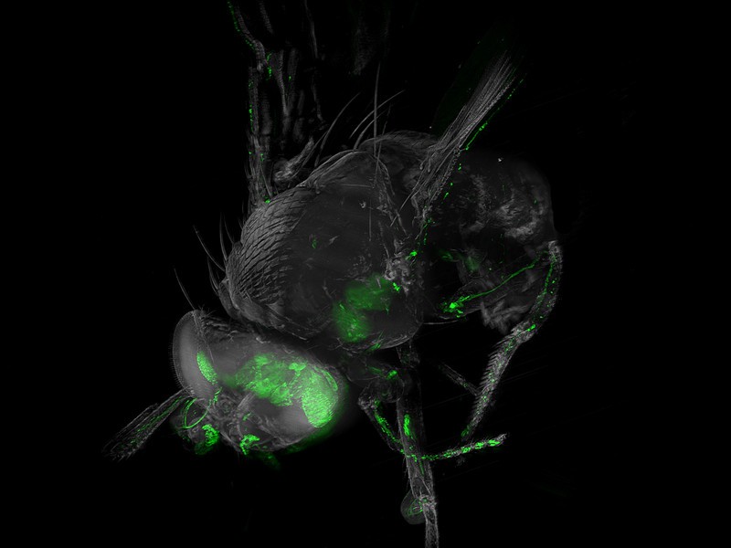
Ultramicroscopy: Brain and optial system of drosophila melanogaster
Advances in cellular microscopy: at TU Wien (Vienna), flies were made transparent, so that individual nerve cells, marked with fluorescent molecules, can be examined directly in the animal. The nervous system of an animal can be studied by cutting it up into thin layers - however this inevitably leads to the destruction of the cellular structures in the tissue. Analyzing complex nerve connections is then hardly possible. The far more elegant method is the so called optical "clearing" of the various tissues using chemical processes that make the animal transparent. Interesting structures in the tissue can be selectively marked and analyzed. At the Vienna University of Technology, a clearing method has now been developed that can be applied to insects, which is a particularly difficult task. With an improved light-sheet microscope (a so-called ultramicroscope), it is now possible to image large nerve tissue samples and take high-resolution pictures of complex neural networks that have been labeled with fluorescent molecules.
TO READ THIS ARTICLE, CREATE YOUR ACCOUNT
And extend your reading, free of charge and with no commitment.