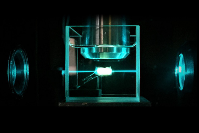
1/3 images In the Ultramicroscope Tumor illuminated by the light sheet Tumor illuminated by the light sheet
1/3 images In the Ultramicroscope Tumor illuminated by the light sheet Tumor illuminated by the light sheet - In order to analyze tumors, they have to be cut into thin slices. Now, a new technology has been developed that makes pieces of the tumor visible in 3D without cutting them. After cancer surgery, the crucial question is: Are there possibly cancer cells left behind that can continue to grow, or has the entire tumor actually been removed? To find out, the tumor is examined by pathologists. Until now, thin sections were made which were then analyzed under a microscope. A new technique, developed at TU Wien (Vienna), together with the TU Munich, could now initiate a revolution in pathology: Tumor tissue is made transparent and illuminated with a special ultramicroscope. This makes it possible to analyze all the tissue removed in 3D without the need for slicing up the tumor. That way, the reliability of the diagnosis can be significantly increased.
TO READ THIS ARTICLE, CREATE YOUR ACCOUNT
And extend your reading, free of charge and with no commitment.