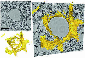
Sections through the three-dimensional reconstruction volume (upper left, grey) around a pulmonary alveolus with hyaline membrane (lower left, yellow). In the centre is the air bubble (alveolus). The electron density is represented by different shades of grey. On the inside of the air bubble is a layer of proteins and dead cell residues, the so-called hyaline membrane. This deposit, which can be represented in its three-dimensional structure for the first time by the new method, reduces the gas exchange and leads to respiratory distress. Photo: Tim Salditt, Marina Eckermann
Sections through the three-dimensional reconstruction volume ( upper left, grey ) around a pulmonary alveolus with hyaline membrane (lower left, yellow ). In the centre is the air bubble (alveolus). The electron density is represented by different shades of grey. On the inside of the air bubble is a layer of proteins and dead cell residues, the so-called hyaline membrane. This deposit, which can be represented in its three-dimensional structure for the first time by the new method, reduces the gas exchange and leads to respiratory distress. Photo: Tim Salditt, Marina Eckermann Researchers led by Göttingen University develop new three-dimensional imaging technique to visualize tissue damage in severe Covid-19 Physicists at the University of Göttingen, together with pathologists and lung specialists at the Medical University of Hannover, have developed a three-dimensional imaging technique that enables high resolution and three-dimensional representation of damaged lung tissue following severe Covid-19. Using a special X-ray microscopy technique, they were able to image changes caused by the coronavirus in the structure of alveoli (the tiny air sacs in the lung) and the vasculature.
TO READ THIS ARTICLE, CREATE YOUR ACCOUNT
And extend your reading, free of charge and with no commitment.