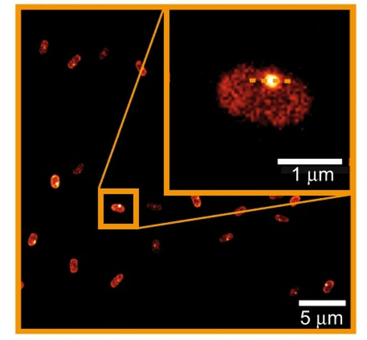
A dormant spore of the bacterium Bacillis subtilis, visualized with the help of the new technique by Brul’s team. Copyright: UvA
A dormant spore of the bacterium Bacillis subtilis, visualized with the help of the new technique by Brul's team. Copyright: UvA - A multidisciplinary team coordinated by biologist Stanley Brul of the University of Amsterdam's Swammerdam Institute for Life Sciences has improved a technique to study live cells in time with the help of fluorescence and rescan confocal microscopy. The technique can be applied to existing microscopy set-ups, making it an easy and cheap solution when higher resolution is needed. The findings were published in Scientific Reports on 24 March. The invention of confocal microscopy and fluorescent markers has made it possible to study live cells and processes within them. Something that used to be impossible with traditional microscopes. And the possibilities continue to increase, both with the development of even better fluorescent markers and improved microscopy techniques.
TO READ THIS ARTICLE, CREATE YOUR ACCOUNT
And extend your reading, free of charge and with no commitment.