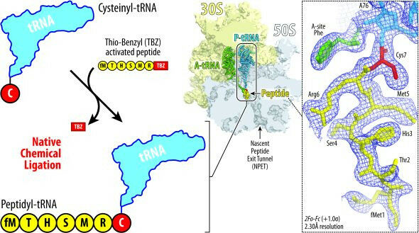
Illustration showing the principle of native chemical ligation approach developed by Syroegin, et al. Addition of the cysteine amino acid (red) to tRNA (blue, top left) allows for the tRNA to fuse to a peptide (yellow, lower left). The resulting ribosome structure (middle) and the captured electron density maps for the peptidyl-tRNA inside the ribosome (right) were obtained by X-ray crystallography in the UIC experiments.
Illustration showing the principle of native chemical ligation approach developed by Syroegin, et al. Addition of the cysteine amino acid ( red ) to tRNA (blue, top left) allows for the tRNA to fuse to a peptide (yellow, lower left). The resulting ribosome structure ( middle ) and the captured electron density maps for the peptidyl-tRNA inside the ribosome ( right ) were obtained by X-ray crystallography in the UIC experiments. Inside tiny cellular machines called ribosomes, chains of genetic material called messenger RNAs (mRNAs) are matched with the corresponding transfer RNAs (tRNAs) to create sequences of amino acids that exit the ribosome as proteins. Unfinished proteins are called nascent chainsm and they are left attached to the ribosome. Scientists know that some of these nascent chains can regulate the activity of the ribosome and that the nascent chains can sometimes interfere with antibiotics - many of which work by targeting bacterial ribosome activity. Scientists do not know why this happens, mainly because it is hard to visualize what the ribosome-peptide-drug interactions look like while the unfinished proteins are still tethered to the ribosome.
TO READ THIS ARTICLE, CREATE YOUR ACCOUNT
And extend your reading, free of charge and with no commitment.