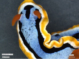
To show that 3D imaging with MIET-SMLM is compatible with biological samples, cells were seeded on a cover glass coated with 10 nm of gold and 5 nm of SiO2 using standard immunofluorescence sample prepa-ration procedure. The artistic rendering illustrates cells imaging on a gold surface resolving microtubules network and clathrin coated pits. Photo: Alexey Chizhik
To show that 3D imaging with MIET-SMLM is compatible with biological samples, cells were seeded on a cover glass coated with 10 nm of gold and 5 nm of SiO2 using standard immunofluorescence sample prepa-ration procedure. The artistic rendering illustrates cells imaging on a gold surface resolving microtubules network and clathrin coated pits. Photo: Alexey Chizhik Research team led by Göttingen University combine two techniques to achieve isotropic super -resolution imaging Over the last two decades, microscopy has seen unprecedented advances in speed and resolution. However, cellular structures are essentially three-dimensional, and conventional super-resolution techniques often lack the necessary resolution in all three directions to capture details at a nanometer scale. A research team led by Göttingen University, including the University of Würzburg and the Center for Cancer Research in the US, investigated a super-resolution imaging technique that involves combining the advantages of two different methods to achieve the same resolution in all three dimensions; this is -isotropic- resolution. The results were published in Science Advances . Despite tremendous improvements in microscopy, there still exists a remarkable gap between resolution in all three dimensions.
TO READ THIS ARTICLE, CREATE YOUR ACCOUNT
And extend your reading, free of charge and with no commitment.