 Single-cell microwell array
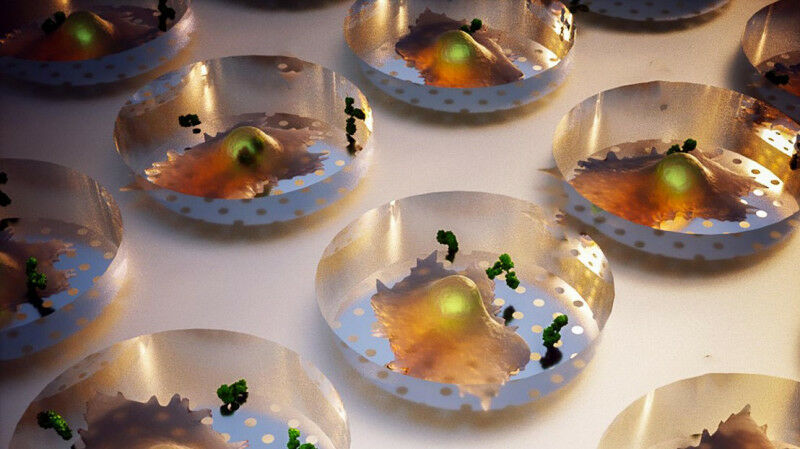
Single-cell microwell array © BIOS EPFL Researchers have used a nanoplasmonics approach to observe the real-time production of cell secretions, including proteins and antibodies; an advancement that could aid in the development of cancer treatments, vaccines, and other therapies. Cell secretions like proteins, antibodies, and neurotransmitters play an essential role in immune response, metabolism, and communication between cells. Understanding cell secretions is key for developing disease treatments, but current methods are only able to report the quantity of secretions, without any detail as to when and where they are produced. Now, researchers in the BIOnanophotonic Systems Laboratory ( BIOS ) in the School of Engineering and at the University of Geneva have developed a novel optical imaging approach that gives a four-dimensional view of cell secretions in both space and time. By placing individual cells into microscopic wells in a nanostructured gold-plated chip, and then inducing a phenomenon called plasmonic resonance on the chip's surface, they are able to map secretions as they are being produced, while observing cell shape and movement. As it provides an unprecedentedly detailed view of how cells function and communicate, the scientists believe their method, recently published in Nature Biomedical Engineering , has "tremendous" potential for pharmaceutical development as well as fundamental research. "A key aspect of our work is that it allows us to screen cells individually in a high-throughput fashion.
TO READ THIS ARTICLE, CREATE YOUR ACCOUNT
And extend your reading, free of charge and with no commitment.
Your Benefits
- Access to all content
- Receive newsmails for news and jobs
- Post ads