 Confocal fluorescence micrographs of cancer cells (BG4, ’H2AX) in the presence of the new photosensitizer DBI. On the left (before irradiation), the nucleus is marked by a fluorescent blue dye, while the photosensitizer DBI is found in fluorescent green vesicles. On the right (after irradiation), DBI has generated reactive oxygen species (ROS) that are highly toxic to the cell. The areas affected appear as fluorescent red dots. Above the micrographs are models of the complex that DBI forms with specific DNA sequences (telomeres).
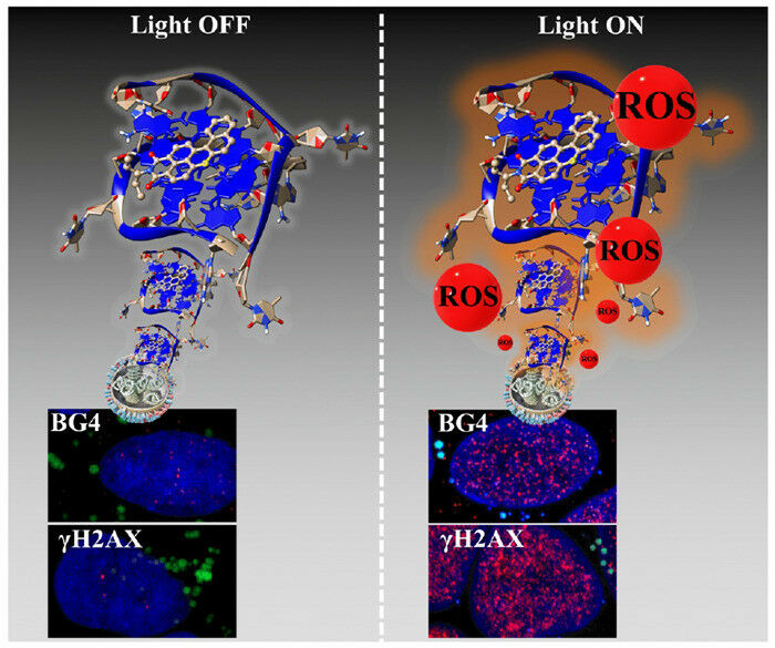
Confocal fluorescence micrographs of cancer cells (BG4, 'H2AX) in the presence of the new photosensitizer DBI. On the left (before irradiation), the nucleus is marked by a fluorescent blue dye, while the photosensitizer DBI is found in fluorescent green vesicles. On the right (after irradiation), DBI has generated reactive oxygen species (ROS) that are highly toxic to the cell. The areas affected appear as fluorescent red dots. Above the micrographs are models of the complex that DBI forms with specific DNA sequences (telomeres). Sabouri et al. Nucleic Acids Research Scientists have designed a molecule that can accumulate in cancer cells specifically and become toxic upon exposure to light.
TO READ THIS ARTICLE, CREATE YOUR ACCOUNT
And extend your reading, free of charge and with no commitment.
Your Benefits
- Access to all content
- Receive newsmails for news and jobs
- Post ads