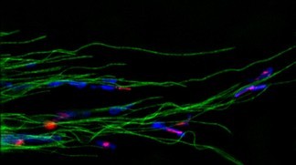
 SiR-tubulin stained in green©Kai Johnsson/EPFL
Cells have their own tiny skeletons that are responsible for many important cellular functions. EPFL scientists have developed novel fluorescent probes for imaging these important structures easily and with unprecedented resolution. Like our own bodies, cells have their own skeletons called 'cytoskeletons' and are made of proteins instead of bones. These network-like structures maintain the cell's shape, provide mechanical support, and are involved in critical processes of the cell's lifecycle. The cytoskeleton is an object of intense scientific and medical research, which often requires being able to observe it directly in cells. Ideally, this would involve highly-fluorescent molecules that can bind cytoskeletal proteins with high specificity without being toxic to the cell. Publishing in Nature Methods , EPFL scientists have exploited the properties of a new fluorescent molecule, also developed at EPFL, to generate two powerful probes for the imaging of the cytoskeleton with unprecedented resolution.
TO READ THIS ARTICLE, CREATE YOUR ACCOUNT
And extend your reading, free of charge and with no commitment.
Your Benefits
- Access to all content
- Receive newsmails for news and jobs
- Post ads