Photo: privat Florian Grüner
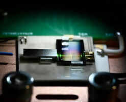
Photo: privat Florian Grüner - How do immune cells move in infectious regions of the body? And how do newly developed agents reach places, for example, where they can fight tumors? X-ray fluorescence imaging, developed further at Universität Hamburg, provides new insight into these questions. Now a team at Universität Hamburg wants to improve access to this technology in cooperation with Siemens- Healthineers and TU Berlin. X-ray fluorescence imaging could be a key application for understanding medical and pharmacological issues. At Universität Hamburg, a team headed by experimental physicist Florian Grüner is conducting research. Despite initial breakthroughs, an unsolved problem remains: to date, the imaging method can be used only in a particle accelerator-based synchrotron plant because only these large facilities are capable of delivering the special x-ray parameters required for imaging. Thus, access to this highly promising imaging is also highly restricted, for example for the Global South. From the large research facility to the small laboratory.
TO READ THIS ARTICLE, CREATE YOUR ACCOUNT
And extend your reading, free of charge and with no commitment.