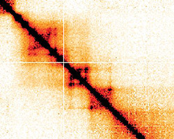
Hi-C contact map
Characterizing chromosome structure is fundamental to a better understanding of gene expression. Current experimental methods helped to build mechanistic models of chromosome folding, however they could not be formally validated so far by independent techniques. This is what the Giorgetti group just did - thanks to a new method they developed to measure chromosome structure quantitatively in living cells. The linear molecules of DNA that constitute a eukaryotic genome have to be carefully organized within the nucleus to be able to correctly direct gene expression. Research has revealed a hierarchical organization into chromosomal territories, compartments, domains, and eventually chromatin loops that serve to bring transcriptional enhancers in proximity of their target promoters. In particular, TADs (Topologically Associating Domains), which were discovered in 2012, are considered a fundamental functional unit in chromosome folding. The current understanding of chromosome folding largely relies on methods based on chromosome conformation capture techniques (3C).
TO READ THIS ARTICLE, CREATE YOUR ACCOUNT
And extend your reading, free of charge and with no commitment.