To play the video, it will be loaded from a Webserver of Google(TM) LLC. Therefore data will be transmitted to Inc. . Google(TM) Privacy Policy Youtube Terms of Service
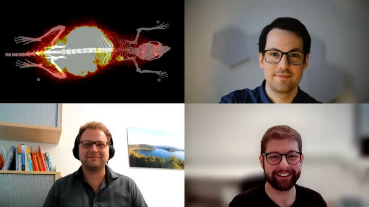
To play the video, it will be loaded from a Webserver of Google(TM) LLC. Therefore data will be transmitted to Inc. Google(TM) Privacy Policy Youtube Terms of Service - Researchers develop imaging methods to examine bodily processes from the individual building blocks to the whole system / Proof-of-principle study on SNAP-tag technology published in the journal "Chemical Communications" Video in English, subtitles available in English and German. Processes and structures within the body that are normally hidden from the eye can be made visible through medical imaging. Scientists use imaging to investigate the complex functions of cells and organs and search for ways to better detect and treat diseases. In everyday medical practice, images from the body help physicians diagnose diseases and monitor whether therapies are working. To be able to depict specific processes in the body, researchers are developing new techniques for labelling cells or molecules so that they emit signals that can be detected outside the body and converted into meaningful images.
TO READ THIS ARTICLE, CREATE YOUR ACCOUNT
And extend your reading, free of charge and with no commitment.