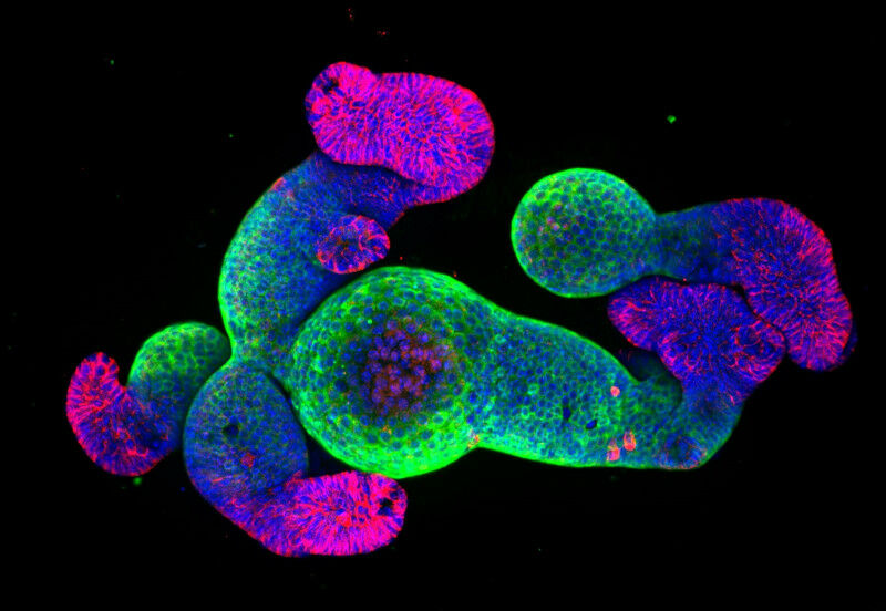
Cells within gut organoids are able to self-organize into structures that mimic intestinal villi (green) and crypts (red). Cells’ nuclei are shown in blue.
Cells within gut organoids are able to self-organize into structures that mimic intestinal villi ( green ) and crypts ( red ). Cells' nuclei are shown in blue. Using miniature guts grown in a dish and 3D biophysical modelling, FMI researchers and their collaborators have uncovered the forces that give the intestinal wall its classic brushlike appearance. The findings can help to understand how the gut takes form during development — and how this process goes awry in disease. Like miniature construction workers, cells orchestrate the building of three-dimensional tissues and organs during embryonic development. To understand the rules governing the growth and repair of these complex structures, FMI group leader Prisca Liberali and her team analyze tiny replicas of organs — 3D clusters of cells known as 'organoids' — that look and function almost like their full-sized counterpart. Gut organoids derived from single cells are able to form an intricate structure organized into fingerlike protrusions called villi and microscopic pits known as crypts.
TO READ THIS ARTICLE, CREATE YOUR ACCOUNT
And extend your reading, free of charge and with no commitment.