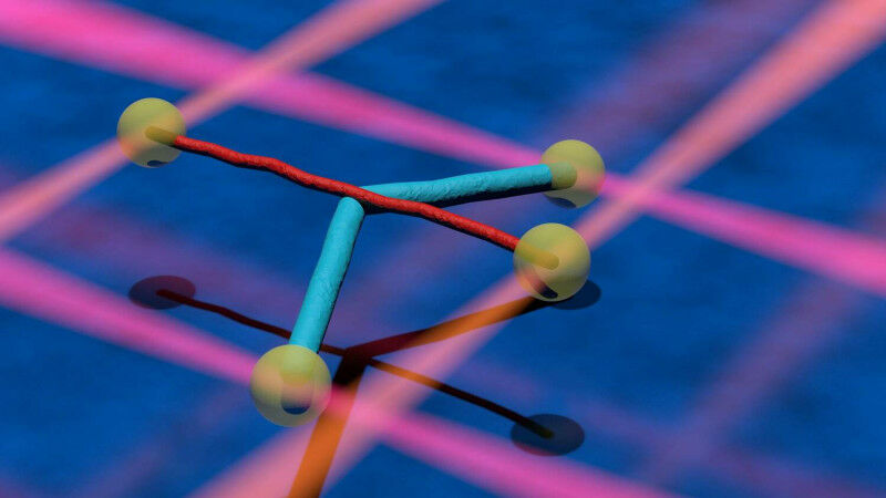
Two different cellular filaments (cyan and red) are attached to microscopic beads (yellow). The particles are held in place with the help of laser beams (orange and red beams). The filaments are crossed and then moved over each other as if playing a violin. They can interact so strongly that one of the filaments is bent (blue). Photo: Markus Osterhoff, Institute for X-ray Physics
Two different cellular filaments (cyan and red) are attached to microscopic beads ( yellow ). The particles are held in place with the help of laser beams (orange and red beams). The filaments are crossed and then moved over each other as if playing a violin. They can interact so strongly that one of the filaments is bent ( blue ). Photo: Markus Osterhoff, Institute for X-ray Physics University of Göttingen research team investigate microtubules Just as the skeleton and muscles move the human body and hold its shape, all the cells of the body are stabilised and moved by a cellular skeleton. Unlike our skeleton, this cellular skeleton is a very dynamic structure, constantly changing and renewing itself. It consists of different types of protein filaments, which include intermediate filaments and microtubules.
TO READ THIS ARTICLE, CREATE YOUR ACCOUNT
And extend your reading, free of charge and with no commitment.