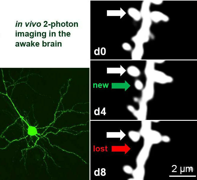
Chronic 2-photon imaging of fluorescently labelled visual cortical neurons in the living adult mouse brain: within the first 4 days of imaging, from day 0 (d0) to day 4 (d4), one dendritic spine (white dendritic protrusion) was newly formed (green arrow); after selective visual stimulation over the next 4 days (until d8), the spine was eliminated (red arrow). White arrow points to a stable spine. Dendritic spine elimination in this paradigm is usually only seen in juvenile mice, but also observed in adult PSD-95 deficient neurons, indicating that PSD-95 is necessary for synapse maturation and stabilization. Photo: S Löwel
Chronic 2-photon imaging of fluorescently labelled visual cortical neurons in the living adult mouse brain: within the first 4 days of imaging, from day 0 (d0) to day 4 (d4), one dendritic spine (white dendritic protrusion) was newly formed (green arrow); after selective visual stimulation over the next 4 days (until d8), the spine was eliminated (red arrow). White arrow points to a stable spine. Dendritic spine elimination in this paradigm is usually only seen in juvenile mice, but also observed in adult PSD-95 deficient neurons, indicating that PSD-95 is necessary for synapse maturation and stabilization. Photo: S Löwel Scientists at Göttingen University discover structural changes in adult mice brains as seen in young animals Understanding the cellular and molecular mechanisms underlying brain -plasticity-(how the brain can learn, develop and reorganise itself) is crucial for explaining many illnesses and conditions. Neurocientists from the University of Göttingen and University Medical Center Göttingen (UMG) have now managed to repeatedly image synapses, the tiny contact sites between neurons, in awake adult mice. They are the first to discover that adult neurons in the primary visual cortex with an increased number of -silent synapses- (ie newly formed synapses that are inactivated), lacking a certain protein (PSD-95), display structural changes that were previously only reported in young mice. This research by the Collaborative Research Centre CRC889 was published in PNAS.
TO READ THIS ARTICLE, CREATE YOUR ACCOUNT
And extend your reading, free of charge and with no commitment.