 TSPO protein (in green) was quantified in microglia (in red) in proximity to lesion characteristic of Alzheimer’s disease, the amyloid plaques (in blue) and pTau lesions (in white), in post mortem human brain samples.
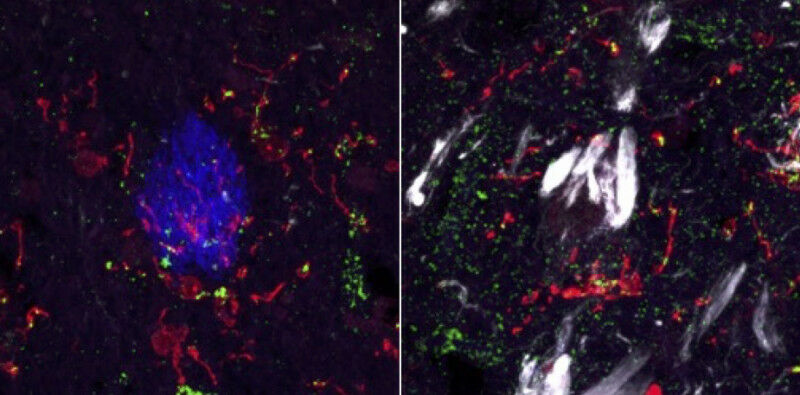
TSPO protein ( in green ) was quantified in microglia ( in red ) in proximity to lesion characteristic of Alzheimer's disease, the amyloid plaques ( in blue ) and pTau lesions (in white), in post mortem human brain samples. Stergios Tsartsalis - An international team co-led by UNIGE and HUG has decoded the only protein that can be used to ''see'' neuroinflammation. This discovery will improve the understanding of neurological and psychiatric disease mechanisms. Inflammation is the sign that our body is defending itself against an aggression. But when this response escalates, for example in the brain, it can lead to serious neurological or psychiatric diseases. A team from the University of Geneva , the University Hospitals of Geneva (HUG), Imperial College London and Amsterdam UMC, investigated a marker protein targeted by medical imaging to visualise cerebral inflammation, but whose interpretation was still uncertain. The team reveals that a large quantity of this protein goes hand in hand with a large quantity of inflammatory cells, but its presence is not a sign of their overactivation.
TO READ THIS ARTICLE, CREATE YOUR ACCOUNT
And extend your reading, free of charge and with no commitment.
Your Benefits
- Access to all content
- Receive newsmails for news and jobs
- Post ads