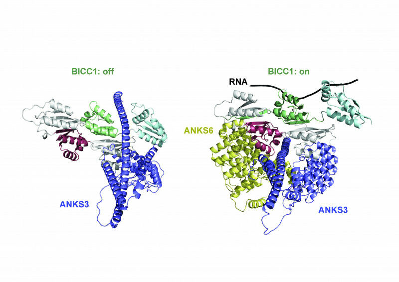
The three proteins, BICC1, ANKS3, and ANKS6 interacting to bind and regulate mRNA in asymmetrical development of organs. Credit: Benjamin Rothé and Zhidian Zhang (EPFL)
The three proteins, BICC1, ANKS3, and ANKS6 interacting to bind and regulate mRNA in asymmetrical development of organs. Credit: Benjamin Rothé and Zhidian Zhang (EPFL) Researchers uncover an intricate protein network controlling asymmetrical development of organs in the embryo, providing insights into genetic disorders and fundamental biology. In order to keep track of their environment, cells use cilia, antenna-like structures that can sense a variety of stimuli, including the flow of fluids outside the cell. Genetic defects that cause cilia to malfunction and lose their sensory abilities can result in disorders known as "ciliopathies", including polycystic kidney diseases; but they can also disrupt the correct asymmetric positioning of internal organs during embryonic development - what is known as "organ laterality". An example of such asymmetry is the heart, which is typically located on the left side, and positioning its blood vessels correctly in a left-right asymmetrical arrangement is critical for efficient oxygen supply around the body. "Therefore, insights into the molecular mechanisms that mediate the sensory functions of cilia to regulate organ laterality are important," says Professor Daniel Constam at EPFL's School of Life Sciences (Swiss Institute for Experimental Cancer Research). In a new study, researchers led by Constam and Professor Matteo Dal Peraro (EPFL Institute of Bioengineering), have found that the factor which is activated by flow-sensing cilia to specify organ laterality is tightly regulated by two other ciliopathy-associated proteins whose molecular functions have been elusive until now.
TO READ THIS ARTICLE, CREATE YOUR ACCOUNT
And extend your reading, free of charge and with no commitment.