Photo credits: Christian Jaeggi for Novartis
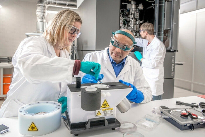
Photo credits: Christian Jaeggi for Novartis - Cryo-electron microscopy, or cryo-EM for short, has revolutionized the way scientists image the smallest of structures. In a short video, the heads of the joint Novartis-FMI cryo-EM center discuss how the technique is advancing biomedical research and drug discovery, and where the field will go in the near future. Cryo-EM is a powerful technique for imaging structures as small as viruses. It involves freezing proteins or other biomolecules in a thin layer of ice, then hitting them with a beam of electrons to determine their 3D shape in stunning detail. These structures can help researchers to understand how medically important proteins work and how drugs could target them. The Novartis campus in Basel is home to a fully operational cryo-EM center, shared between Novartis and the FMI. The center began operating in November 2016.
TO READ THIS ARTICLE, CREATE YOUR ACCOUNT
And extend your reading, free of charge and with no commitment.