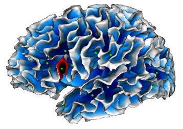
 © SCALP team / INT Mapping of cortical fold depths. In green: sulcal pits (the deepest point of each fold). In red: localization of the abnormality detected in autistic children .
Scientists at CNRS, Aix-Marseille Université and AP-HM have identified a cerebral marker specific to autism that can be detected by MRI and is present as from the age of two years. The abnormality thus detected consists in a less deep fold in Broca's area, a region of the brain specialized in language and communication, functions that are impaired in autistic patients. This discovery may assist in the earlier diagnosis and management of these patients. It has been made possible by the medical imaging processing skills of the Institut de Neurosciences de la Timone (CNRS/Aix-Marseille Université) and access to a homogeneous cohort of patients diagnosed at a very young age and all assessed using the same protocol at the Centre de Ressources Autisme PACA. The results of their collaboration are published on 12 January 2016 in Biological Psychiatry: Cognitive Neurosciences and Neuroimaging . The autistic spectrum covers a range of neuro-developmental disorders (typical autism, Asperger's syndrome or pervasive developmental disorders not otherwise specified) which mainly affect social relationships and communication. These disorders are associated with abnormal development of the brain.
TO READ THIS ARTICLE, CREATE YOUR ACCOUNT
And extend your reading, free of charge and with no commitment.
Your Benefits
- Access to all content
- Receive newsmails for news and jobs
- Post ads