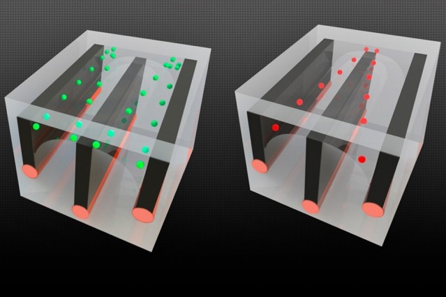
By integrating conductive wires along with microfluidic channels in long fibers, the researchers were able to demonstrate the ability to sort cells - in this case, separating living cells from dead ones, because the cells respond differently to an electric field. The live cells, shown in green, are pulled toward the outside edge of the channels, while the dead cells (red) are pulled toward the center, allowing them to be sent into separate channels. Illustrations courtesy of the researchers.
Fibers containing systems for mixing, separating, and testing fluids may open up new possibilities for medical screening. Microfluidics devices are tiny systems with microscopic channels that can be used for chemical or biomedical testing and research. In a potentially game-changing advance, MIT researchers have now incorporated microfluidics systems into individual fibers, making it possible to process much larger volumes of fluid, in more complex ways. In a sense, the advance opens up a new "macro" era of microfluidics. Traditional microfluidics devices, developed and used extensively over the last couple of decades, are manufactured onto microchip-like structures and provide ways of mixing, separating, and testing fluids in microscopic volumes. Medical tests that only require a tiny droplet of blood, for example, often rely on microfluidics. But the diminutive scale of these devices also poses limitations; for example, they generally aren't useful for procedures that need larger volumes of liquid to detect substances present in minute amounts.
TO READ THIS ARTICLE, CREATE YOUR ACCOUNT
And extend your reading, free of charge and with no commitment.