Pixabay. Credit: Darko Stojanovic
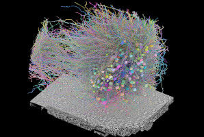
Scientists at UCL have designed a virtual modelling technique which can create highly detailed 3D models of individual cancerous tumours and simulate the delivery of drugs in order to predict their effectiveness. In the study, researchers acquired high-resolution images of surgically-resected tumours and used mathematical modelling to run detailed computational experiments. This allowed them to study the transport of blood, biological fluids and drugs, and their complex interactions with tissue. Their new technique, named REANIMATE (REAlistic Numerical Image-based Modelling of biologicAl Tissue substratEs) enables researchers to visualise and interact with large, 3D, virtual models of tumour tissue samples and treat them as living specimens. This will enable scientists to perform complex computational experiments to generate new insights into how individual tumours react to specific treatments. In the study researchers used optical imaging of extracted tumour tissue that had been rendered transparent using a cocktail of chemical treatments. These can show fine detail such as blood vessel networks and cell nuclei, which can be seen across entire organs at very high resolution by using fluorescently-labelled probes that bind to specific structures.
TO READ THIS ARTICLE, CREATE YOUR ACCOUNT
And extend your reading, free of charge and with no commitment.